Thorax

Chest CT scan
A chest CT scan can be performed without any problems if the patient's condition allows for a brief pause in breathing (up to 20 seconds). For most patients, this is not a problem at all.
A chest CT scan allows for the visualization of all structures within the chest; through the use of filters and contrast dye, many abnormalities can be visualized more clearly than with X-rays.
CASE: Cindy, a 7-year-old Malinois, suddenly experiencing shortness of breath
Cindy was referred due to shortness of breath that developed quite rapidly.
Clinical examination: normal temperature, pulse = 120, respiration: panting/short of breath.
Right lung field: significantly diminished breath sounds; left lung field: no abnormalities. Mucous membranes pink; lymph nodes not enlarged.
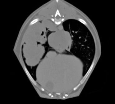
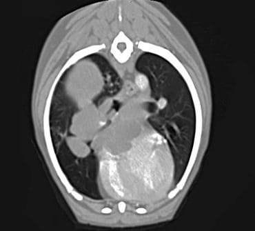
The referring veterinarian suspected lung lobe torsion. According to the radiologist, the CT images “showed a multilobular tumor in the right lung with invasion into the right atrium, the vena cava, and the right main bronchus. A mass is also visible in the right chest wall.” The banner at the top of this page displays several CT images. The differential diagnosis may include lymphoma, a histiocytic sarcoma, or a primary lung tumor.
Cindy was euthanized after a few days.