General Surgery
Fully equipped operating rooms
The clinic has three fully equipped operating rooms, fitted with state-of-the-art anesthesia and monitoring equipment. To ensure safe anesthesia, a thorough examination of the animal scheduled for surgery is conducted beforehand.
Aftercare
On the day of your pet’s surgery, he or she will still be a little groggy from the anesthesia. Please leave him or her alone as much as possible, and do not give him or her any food. Water is okay, but not too much—give small amounts at a time. The day after the surgery, he or she can eat and drink normally again.
Please don't hesitate to call if your pet refuses to eat or drink in the coming days, or if there is noticeable swelling around the wound. Blood loss is also a reason to contact us.
The surgical incision has been closed with stitches; these should be removed after 8 to 10 days (you will be told after the surgery exactly how many days). You can come in during one of our office hours to have them removed. To prevent your pet from licking the wound, it will be wearing an Elizabethan collar or a special protective suit. The collar or suit must remain on, because licking can quickly cause surgical wounds to become infected, which can cause the stitches to come loose.
Unfortunately, we must charge you for any treatment costs resulting from the animal licking the wound. Dogs may be walked on a leash, but cats must remain indoors until the stitches have been removed.
What types of surgeries do we offer? Here is an overview:
THE HEAD AND NECK:
The auricles (ears):
• Surgical treatment of an othematoma or bloody ear
• Suturing wounds on the auricles
• Removal of growths on the auricles, e.g., partial or total auricle amputations.
The ear canals (ears):
• Partial (Zepp procedure) or total (TECA = total ear canal ablation) removal
• Removal of an ear canal with or without lateral bulla osteotomy
• Ventral bulla osteotomy
• Removal of benign masses from the ear canal, e.g., a polyp in cats
The eyes and eyelids:
• Cosmetic removal of eyelid cysts (most commonly used method: H-plasty)
• Surgical treatment of entropion (inward turning of the eyelids)
• Enucleation of the eyeball, or the complete removal of the eyeball in cases of irreversible damage such as glaucoma or tumors.
• Surgical treatment of a cherry eye (primarily in young dogs)
The Nose (Mirror):
• Removing growths or suturing wounds
• Widening the nostrils in short-snouted breeds (brachycephalic breeds)
The throat region:
• Shortening the soft palate (palatum molle) in brachycephalic breeds
• Removing masses from the nasopharynx
The windpipe or trachea:
• Removal of masses or foreign objects from the cervical trachea
• Suturing wounds on the cervical trachea
The salivary glands:
• Removal of affected salivary glands and drainage of salivary gland cysts (mucoceles)
The Thyroid Glands:
• Removal of a thyroid tumor in dogs
The lips and tongue:
• Removal of growths or suturing of wounds.
• Partial tongue amputation
THE ABDOMINAL CAVITY:
The liver
• Taking surgical biopsies and removing tumors whenever possible
The spleen:
• In general, the spleen is completely removed when abnormalities are found (splenectomy)
The stomach:
• Taking surgical biopsies
• Removing masses or foreign objects (gastrotomy)
• Preventive (large breeds with deep chests) or curative fixation of the stomach (gastropexy)
The small intestine:
• Taking surgical biopsies (through the entire wall thickness)
• Removing foreign objects from the small intestine (enterotomy)
• Removing a section of the intestine in cases of severe damage caused by a lodged foreign object or tumors (enterectomy)
• Surgical treatment of intussusception
The large intestine:
• Removal of dried-up material that can accumulate after eating bones (colotomy)
• Removal of masses from the large intestine (colectomy)
• Surgical fixation of the large intestine, e.g., in cases of rectal prolapse or perineal hernia (colopexy)
• Surgical treatment of megacolon in cats (partial or total colectomy)
The kidneys:
• Taking surgical biopsies
• Removing an entire affected kidney (nephrectomy)
The bladder:
• Removal of bladder stones and tumors
• Partial cystectomies for tumors.
The urinary tract in male animals:
• Performing a penectomy on a male cat
• Performing a urethrostomy on a male dog
• In both cases, the animals are provided with a so-called “new urinary opening” when they can no longer urinate due to a blockage caused by stones or grit.
The male reproductive system:
• Normal castration of a male dog
• Castration of a male dog with one or both testicles in the groin
• Castration of a male dog with one or both testicles in the abdominal cavity
• When one or both testicles have not descended, this is referred to as cryptorchidism.
• Draining or (partially) removing prostate cysts or abscesses
• Suturing wounds or removing masses on the penis or prepuce
• Surgical treatment of urethral prolapse in male dogs
The reproductive system of female animals:
• Standard spaying of a young female dog (removal of the ovaries only)
• Laparoscopic spaying of a young female dog
• Complete removal of the uterus and ovaries in older female dogs in cases of uterine infection (pyometra), cysts, or tumors on the ovaries.
• Suturing wounds and removing masses in the vagina and on the vulva
Mammary Glands:
• Total (complete mastectomy) or partial removal of the mammary gland in cases of tumors in the mammary glands.
THE EXTREMITIES:
The Limbs
• Suturing wounds and removing growths.
The tail:
• Suturing wounds, removing growths (sebaceous cysts), and performing a (partial) tail amputation in cases of large defects.
THE ABDOMINAL AND THORACIC WALLS:
• Suturing wounds and removing growths
HERNIAS OR FRACTURES:
• Surgical treatment of all types of hernias, such as umbilical hernias (hernia umbilicalis), inguinal hernias (hernia inguinalis), incisional hernias, hernias resulting from trauma, and perineal hernias in male dogs.
THE ANAL REGION:
• Surgical removal of the anal sacs or anal glands in cases of recurrent overfilling or inflammation
• Removal of anal sac tumors
• Removal of perianal growths
ORTHOPEDICS:
• Anterior cruciate ligament rupture
• Patellar luxation
• Shoulder surgery
• Femoral head resections
• For spinal surgery (including herniated disc surgery), we refer patients to a veterinary clinic with extensive expertise in this field.
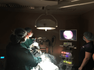
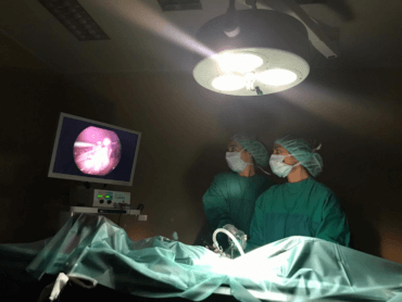
Laparoscopic sterilization
During the spaying of a female dog, normally only the ovaries are removed. If the uterus is abnormal, it may be necessary to remove the entire uterus. In the “traditional” method, an incision is made in the abdominal wall, and the veterinarian will manually ligate and remove the ovaries. The female dog will then no longer go into heat.
Nowadays, it’s also possible to spay a female dog using a minimally invasive procedure (laparoscopy).
Through three small incisions of about 5 mm, we can insert a camera and a few instruments into the abdomen. Using a monitor and the camera’s magnification, the veterinarian has a clear view to assess the organs and the uterus. If everything is in order, the ovaries are removed using electrocautery.
THE BENEFITS OF LAPAROSCOPIC STERILIZATION:
- There is less pain after surgery because no large incision needs to be made. Also, the ovarian ligaments are not pulled.
- The risk of wound infection is significantly lower given the small holes that are made
- There is a lower risk of post-operative bleeding, which is checked immediately using the camera.
- Faster recovery and a quicker return to normal; they’ll start eating sooner and will be allowed to move around more as early as the day after surgery.
- A clear view of all the organs during the operation.
THE DISADVANTAGES OF LAPAROSCOPIC STERILIZATION:
- If the veterinarian determines that the uterus does not look healthy, it will be necessary to perform the surgery using the “traditional” method after all and remove the entire uterus.
- The costs are higher because special equipment is used.