Orthopedic Surgery
Torn Anterior Cruciate Ligament in Dogs
A torn anterior cruciate ligament is one of the most common knee problems in dogs. It is often caused by gradual wear and tear of the ligament. Dogs with a torn anterior cruciate ligament are often in a lot of pain and limp severely. The cruciate ligament ensures that the knee remains stable while walking. The instability that develops in the knee joint after the cruciate ligament tears can also cause the meniscus to become damaged or torn. In the long term, severe wear and tear of the articular cartilage (osteoarthritis) may also develop. If a torn cruciate ligament is not surgically repaired, the dog will remain in pain and lame for the rest of its life.
Making the Diagnosis
The diagnosis is made through a physical examination. During this examination, we perform special tests, such as the drawer test and the tibial compression test. Sometimes these tests can only be performed reliably under light sedation, because the dog may be tense or in pain. In cases of doubt, we also take X-rays to support the diagnosis.
The art strap (Flo rein)
For smaller dogs weighing less than 15 kg, we often use the so-called synthetic ligament technique, also known as the Flo-strap. This technique mimics the function of the torn cruciate ligament using a sturdy strap placed outside the knee joint. This stabilizes the knee so the dog can move properly again. This technique is not suitable for larger dogs because the synthetic ligament is not strong enough for them.
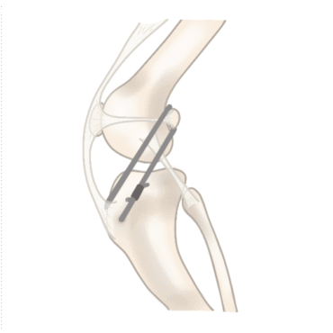
Schematic representation of the Flo rein
The TTA Rapid Method
The TTA Rapid procedure (Tibial Tuberosity Advancement) is a modern surgical technique that restores stability to the knee. By making a small adjustment to the tibia using a special implant, the forces within the knee joint are rebalanced. This adjustment restores stability to the knee during movement. We use this method for dogs weighing approximately 15 kg or more. After recovery, the dog can move actively and pain-free again.
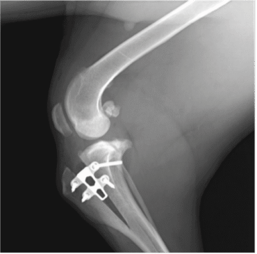
X-ray after TTA surgery
The Surgery, Step by Step
- Your dog will be put under general anesthesia.
- X-rays are taken to accurately determine the necessary correction and the appropriate implant size.
- The dog is then prepared for surgery. The leg is shaved and washed.
- Next, the knee joint is opened: the remnants of the torn cruciate ligament are removed, and the meniscus is examined for damage. If necessary, the damaged portion of the meniscus is removed.
- For smaller dogs, a synthetic ligament is implanted.
- In larger dogs, the tibia is cut in a controlled manner. The “cage” (the implant) is then inserted. It is secured with 4 to 6 screws (depending on the size of the cage).
- Finally, the wound is carefully closed.
After the surgery
Your dog can usually go home the same day. Proper aftercare is very important: exercise should be gradually increased in a gentle and controlled manner. We’ll provide you with detailed aftercare instructions to ensure the recovery goes as smoothly as possible!
Patellar Dislocation
What is patellar dislocation?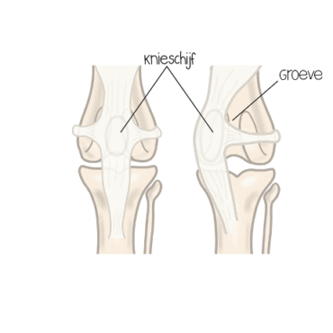
Patellar luxation is a common orthopedic condition in dogs. In this condition, the kneecap (patella) slips out of the groove in the thighbone, where it normally sits. This can occur toward the inside (medial luxation) or the outside (lateral luxation). The condition is often caused by an abnormal alignment of the hind leg or by a groove in the femur that is too shallow. Patellar luxation is common in small dog breeds, but it can also occur in medium and large breeds. In most cases, the luxation occurs inward (medial luxation).
Dogs with patellar luxation can exhibit a variety of symptoms. Sometimes you’ll see a dog suddenly lift a hind leg while walking and “hop” for a moment, before resuming its normal gait. In more severe cases, dogs may continue to limp, experience pain, and develop wear and tear (osteoarthritis) in the knee joint. The severity of the symptoms depends on the degree of the luxation. Because the condition is often chronic and progressive, it is important to make a timely diagnosis and consider appropriate treatment.
Making a Diagnosis
Patellar luxation is diagnosed through a physical examination, during which the veterinarian manually moves the kneecap to determine whether it slips out of its groove. There are several degrees of patellar luxation:
- Grade I: The kneecap can be manually pushed out of its groove, but it springs back into place on its own.
- Grade II: The kneecap frequently pops out of its groove on its own, but can also pop back into place.
- Grade III: The kneecap is often displaced to the side of the groove and remains there, but can be repositioned by applying pressure.
- Grade IV: The kneecap is permanently displaced to the side of the groove and cannot be repositioned.
Surgical Technique
Patellar luxation is usually treated with surgery. The exact steps the veterinarian takes depend on the severity of the condition. Often, several techniques are combined to restore the kneecap to its proper, stable position.
- Deepening the groove: The “groove” in the femur where the patella is supposed to slide is made slightly deeper. This helps keep the patella in place.
- Repositioning the patellar tendon attachment: The point where the tendon attaches to the shinbone is moved slightly so that the pulling force runs straight through the groove again. This helps prevent the kneecap from being pulled out of alignment.
- Tightening the soft tissues: The ligaments and fascia around the knee can be strengthened or tightened slightly to provide additional support.
Combining these techniques makes the knee more stable and greatly increases the chances that the dog will be able to walk pain-free again.
Aftercare
Proper aftercare is essential for a successful recovery. The dog will be sent home with pain medication. It is important that the dog gets plenty of rest during the first few weeks and only goes on short, leashed walks. Climbing stairs, jumping, and playing should be avoided as much as possible.
After a few weeks, the exercise load can be gradually increased. Physical therapy can support recovery and promote muscle building. A follow-up visit to the veterinarian is scheduled to assess whether healing is progressing well. For most dogs, the prognosis after surgery for patellar luxation is very good.
Please contact us for more information
Dierenkliniek den Heuvel here for you 365 days a year!
Contact us