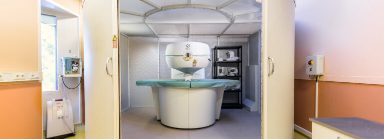
MRI
(Magnetic Resonance Imaging)
What is an MRI?
MRI (Magnetic Resonance Imaging) is an imaging technique that can provide highly valuable information about all kinds of structures in the body that cannot be visualized using other imaging techniques (X-rays, ultrasound). This includes highly detailed images of the brain and spinal cord, as well as cartilage, bones, various abdominal organs, ligaments, and tendons in and around joints, and so on. Furthermore, MRI makes it possible to clearly distinguish between normal and diseased tissue. All of this is achieved without the use of harmful radiation (as is the case with X-rays and CT scans)!
Simply put, an MRI scanner works as follows:
The animal is placed in a strong magnetic field. This causes all the hydrogen nuclei (which are essentially tiny magnets) in the body to align in the same direction. A radio pulse briefly flips the nuclei out of that direction, after which they return to the same direction. In doing so, they emit a small radio signal. This signal is picked up by a coil-shaped antenna surrounding the body and translated into an image by the computer. The speed at which the hydrogen nuclei return to their initial position depends on the type of tissue, creating differences in contrast between the various tissues, which allows us to distinguish them from one another in the image.
A large number of images are taken of the body part being examined, as if it were being sliced into three-dimensional sections. Together, all these images provide a wealth of information that cannot be visualized in any other way.
Safety
Although MRI technology is safe for both humans and animals, there are still a few things to keep in mind:
The MRI scanner contains a very strong magnet. This means you must not come near the scanner while wearing a watch (it will stop immediately), carrying debit or credit cards (they will be erased immediately), having implants in your body (pacemaker, insulin pump, artificial joint, etc.), or wearing certain hearing aids—in short, if you are carrying any metal objects that could be affected by the strong magnetic field. In practice, this means we will ask you to remain outside the MRI examination room.
You can wait in the waiting room—with a cup of coffee, of course.
Why an MRI?
Any owner who takes a sick animal to the vet is, in principle, interested in only three things:
- What's wrong with him? (Diagnosis)
- What are the chances of recovery? (Prognosis)
- What can be done about it? (Therapy)
It should be clear that questions two and three cannot be answered properly without a diagnosis.
It is still quite common that a definitive diagnosis cannot be made using standard diagnostic techniques such as X-rays and ultrasound. Damage to ligaments and tendons, as well as disorders of the brain and spinal cord, are usually not visible on X-rays or ultrasound images. An MRI, however, usually does provide a diagnosis. Just as with people who have “unexplained” symptoms, for many animals a diagnosis is only reached after an MRI, after which targeted treatment and a good prognosis can be provided!
Anesthesia
If you schedule an MRI exam with us, this means your pet will need to be anesthetized. During the exam, which typically takes about three-quarters of an hour, your pet must not move at all.
Proper preparation for a procedure under anesthesia begins at home. The day before the procedure, your pet must not eat anything after 6:00 p.m. Water is allowed, however! The next day, please arrive at the clinic at the scheduled time. Please walk your dog beforehand!
When you bring your pet to us, you do so with the (justified) expectation that he or she will receive the best possible medical care. That is why we always perform a physical examination before the animal is put under anesthesia. Of course, we pay particular attention to the heart and lungs during this examination.
However, a physical examination alone does not reveal all possible issues. That is why it may be important to perform a pre-anesthetic blood test. This may be necessary especially for older animals and those with impaired kidney or liver function. If you are visiting us on referral from another veterinarian, please bring your pet’s medical history (including any blood test results and/or X-rays).
Just as in human medicine, anesthetics used in veterinary medicine are very safe. The risks of anesthesia are therefore minimal for a healthy animal. However, if there are any health issues (unknown to you or us), anesthesia may cause certain problems.
If the pre-anesthesia examination reveals no abnormalities, our team can safely anesthetize your animal and begin the examination. The animal is continuously monitored during anesthesia.
After the examination is complete, the animal is woken up and, as a rule, can go home immediately.
After the investigation
Once the examination of the animal is complete, the results and the images obtained must be evaluated. Often, a diagnosis can be made immediately after the examination. If necessary, images are submitted for further evaluation to experts in the field of medical imaging: Dr. I. Gielen and Prof. H. van Bree of Ghent University.
If you have been referred to us by your own veterinarian, you will be referred back to them, and your veterinarian will receive a report from us as soon as possible (by phone, mail, fax, or email), so that once you are back home, you can work with him or her to develop a plan for further treatment.
You will receive a CD containing all the images from the examination. Of course, we will store and retain all the data.
Syringomyelia
Chiari-like malformation and syringomyelia syndrome is a condition that occurs in a number of small dog breeds. It is best known in the Cavalier King Charles Spaniel, because a large number of Cavalier breeders have their breeding dogs screened for this condition. However, there is no doubt that the condition occurs in a significant number of breeds, possibly even at a higher frequency than in the Cavalier King Charles Spaniel. These include breeds such as the Chihuahua, Pomeranian, Petit Griffon, Pug, and King Charles Spaniel.
This condition can only be diagnosed via MRI. An extensive screening program is currently underway to assess the prevalence of CM/SM (for now, only in Cavaliers).
Thanks to consistent screening for this condition, there has been a clear decline in the number of cases among pedigreed Cavaliers in recent years. Unfortunately, we do not see this trend among the so-called “look-alikes”—dogs sold as Cavaliers without a pedigree.
Dog owners who wish to have their pet(s) examined can make an appointment. We offer a significantly reduced rate for CM/SM screening. The images we produce are evaluated by Dr. P.J.J. Mandigers, a veterinary neurologist.
When to use an MRI and when to use a CT scan?
For some conditions, an MRI is the best way to reach a diagnosis; for others, a CT scan is.
Click HERE to see an overview of which research method is best.