Brain
CT or MRI?
CT provides a clear and, above all, rapid assessment of the skull and surrounding structures. CT can be used to detect skull trauma, extracranial tumors and abscesses, as well as large brain tumors and cerebral hemorrhages.
However, for a thorough brain examination, MRI is the best diagnostic method. This test can be used to detect conditions such as cerebral edema, tumors, encephalitis, meningitis, hematomas, strokes, hydrocephalus, and Chiari malformation/syringomyelia.
CASE 1: Puck, an Airedale Terrier, 9 years old, cannot swallow
Puck, a 9-year-old Airedale terrier, belonged to one of our employees.
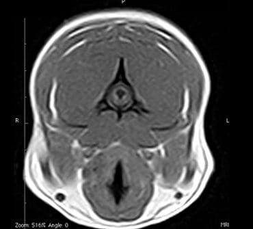
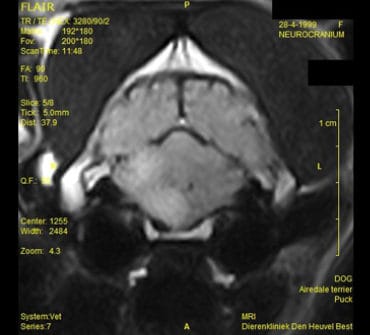
Puck’s symptoms began with what appeared to be excessive drooling. At first, eating and drinking were still going well. An examination of the mouth and throat revealed nothing unusual. Very gradually, the drooling got worse, and at one point it became noticeable that it took a long time for her to slurp a bowl of water dry. Eating also slowed down at that point. There were clearly problems with chewing and swallowing, and when a follow-up examination of the mouth and throat revealed nothing, it was decided to perform an MRI scan of the head.
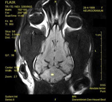
This examination revealed the presence of a tumor in the right half of the pons and medulla oblongata. The tumor’s location in the area where both the trigeminal nerve and the hypoglossal nerve originate explains the patient’s symptoms.
Shortly after the MRI scan, Puck was euthanized because she was virtually unable to swallow. No autopsy was performed, so tissue typing could not be done. In terms of differential diagnosis, a glioma should be strongly suspected.
CASE 2: Boy, a 4-year-old Maltese mix with itchy ears and neck
Boy was referred to us because he had constant itching in his ears and on his neck. He had already been treated with ear ointment, but without success. Because he was also showing signs of pain and frequently rubbing his muzzle on the floor, his veterinarian suspected syringomyelia, a condition most commonly associated with the Cavalier King Charles Spaniel but also found in quite a few other small breeds.
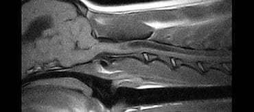
- The MRI scan revealed the following abnormalities:
- Dilation of the lateral cerebral ventricles
- Dilation of the third and fourth ventricles
- Hypoplasia of the caudal fossa, resulting in:
- Herniation of cerebellar tissue through the foramen magnum (Chiari-like malformation)
- Syringomyelia of the cervical spinal cord
After the diagnosis was made, Boy was referred back to his own veterinarian to follow the Rusbridge treatment protocol. After some “trial and error,” he ultimately responded very well to gabapentin.