Neck and Back

Spinal disorders
We see spinal problems in all breeds, from large to small. The most commonly diagnosed condition is herniated nucleus pulposus (HNP). In addition, we regularly encounter extradural or intramedullary tumors, fibrocartilaginous emboli, discospondylitis, or hematomas.
Patients with spinal problems often present with neurological deficits ranging from mild paresis to complete paralysis. In addition, the animals are often in a great deal of pain.
A suspected HNP combined with neurological deficits is a medical emergency! In any case, a high dose of methylprednisolone succinate (MPS) (e.g., Soludeltacortef) must be administered as soon as possible. Other medications are not very effective. DO NOT give NSAIDs as pain relievers: when combined with MPS, this can lead to severe gastrointestinal symptoms! It is important to establish a diagnosis as quickly as possible so that any necessary surgical intervention can be arranged immediately.
Many spinal problems can be diagnosed using a CT scan. However, there are also quite a few conditions that can only be diagnosed with an MRI.
CASE 1: Fred, a 6-year-old Cairn Terrier, with posterior paresis, particularly on the left side
Fred, a 6-year-old Cairn Terrier, was referred to us with signs of paralysis, particularly on the left hind side. Postural reflexes were absent on the left hind side and delayed on the right; spinal reflexes were normal. Palpation of the spine was painful in the thoracolumbar region.
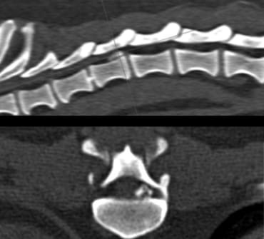
A CT scan of the spine revealed a calcified mass of nucleus material, extradural, on the left side of the spinal canal (see banner images).
After being diagnosed with a herniated disc at L1-L3, the dog was referred, at the request of the referring veterinarian, to a nearby clinic with extensive expertise in spinal surgery. Fred underwent surgery there and made a quick recovery.
CASE 2: Milou, a 4-year-old female Briard with acute, severe neck pain
This dog had been whimpering in pain for two days, keeping her head and neck lowered. Moving her neck was extremely painful. A neurological examination revealed no abnormalities other than the neck pain.
Milou was referred on suspicion of a herniated intervertebral disc (HNP).
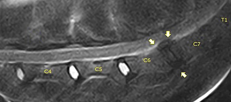
A CT scan of this dog’s neck did not yield a diagnosis: the CT image was normal. Therefore, an MRI of the neck was performed, which confirmed the referring veterinarian’s suspicion: a herniated intervertebral disc (HNP) at the C6-C7 level.
Since Milou showed no neurological deficits and responded well to pain medication, her own veterinarian decided to treat this herniated disc conservatively, after which she made a full recovery.